Anatomy
Head and Neck
The lateral rectus muscle acts to produce which of the following eyeball movements:
Answer:
The lateral rectus muscle acts to abduct the eyeball.Orbital Muscles
Anatomy / Head and Neck / Orbit and Eye
Last Updated: 4th November 2019
Movements of the Eye
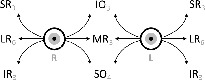
The six extraocular muscles are responsible for turning or rotating the eye about its vertical, horizontal, and anteroposterior axes.
Table: Movements of the Eyeball
| Action | Description | Primary Muscle(s) |
|---|---|---|
| Elevation | Moving pupil superiorly | Superior rectus and inferior oblique |
| Depression | Moving pupil inferiorly | Inferior rectus and superior oblique |
| Abduction | Moving pupil laterally | Lateral rectus |
| Adduction | Moving pupil medially | Medial rectus |
| Medial rotation (intorsion) | Rotating upper part of pupil medially towards nose | Superior oblique |
| Lateral rotation (extorsion) | Rotating upper part of pupil laterally towards temple | Inferior oblique |
Orbital Muscles
ORIGIN:
The recti muscles all originate as a group from a common tendinous ring at the apex of the orbit and form a cone of muscles as they pass forward to their attachment on the eyeball.
![By OpenStax College [CC BY 3.0], via Wikimedia Commons](https://primary-cdn.frcemsuccess.com/wp-content/uploads/2016/07/1412_Extraocular_Muscles-1024x394.jpg)
Structure of the Orbital Muscles. (Image by OpenStax College [CC BY 3.0], via Wikimedia Commons)
ACTION AND INNERVATION:
Table: Overview of the Orbital Muscles
| Extraocular Muscle | Innervation | Function | Clinical Assessment (direction to move eye when testing muscle) |
|---|---|---|---|
| Superior rectus | Oculomotor nerve | Elevation, adduction and medial rotation of eyeball | Look out and up |
| Inferior rectus | Oculomotor nerve | Depression, adduction and lateral rotation of eyeball | Look out and down |
| Medial rectus | Oculomotor nerve | Adduction of eyeball | Look in (in horizontal plane) |
| Lateral rectus | Abducens nerve | Abduction of eyeball | Look out (in horizontal plane) |
| Superior oblique | Trochlear nerve | Depression, abduction and medial rotation of eyeball | Look in and down |
| Inferior oblique | Oculomotor nerve | Elevation, abduction and lateral rotation of eyeball | Look in and up |
ASSESSMENT:
To test the muscles in isolation, the patient can be asked to move their eyeball in certain directions. A lateral position of the eyeball is necessary for testing the inferior and superior recti, whereas a medial position is necessary for testing the inferior and superior oblique. This first movement (laterally or medially) brings the axis of the eyeball into alignment with the axis of the muscle. If the extraocular muscle being tested is paralysed, the patient will be unable to perform the movement and will complain of diplopia.

Clinical Testing: Direction to Move Eye when Testing Muscles (Image by Au.yousef [CC BY-SA 4.0 (https://creativecommons.org/licenses/by-sa/4.0)], from Wikimedia Commons)
Report A Problem
Is there something wrong with this question? Let us know and we’ll fix it as soon as possible.
Loading Form...
- Biochemistry
- Blood Gases
- Haematology
| Biochemistry | Normal Value |
|---|---|
| Sodium | 135 – 145 mmol/l |
| Potassium | 3.0 – 4.5 mmol/l |
| Urea | 2.5 – 7.5 mmol/l |
| Glucose | 3.5 – 5.0 mmol/l |
| Creatinine | 35 – 135 μmol/l |
| Alanine Aminotransferase (ALT) | 5 – 35 U/l |
| Gamma-glutamyl Transferase (GGT) | < 65 U/l |
| Alkaline Phosphatase (ALP) | 30 – 135 U/l |
| Aspartate Aminotransferase (AST) | < 40 U/l |
| Total Protein | 60 – 80 g/l |
| Albumin | 35 – 50 g/l |
| Globulin | 2.4 – 3.5 g/dl |
| Amylase | < 70 U/l |
| Total Bilirubin | 3 – 17 μmol/l |
| Calcium | 2.1 – 2.5 mmol/l |
| Chloride | 95 – 105 mmol/l |
| Phosphate | 0.8 – 1.4 mmol/l |
| Haematology | Normal Value |
|---|---|
| Haemoglobin | 11.5 – 16.6 g/dl |
| White Blood Cells | 4.0 – 11.0 x 10⁹/l |
| Platelets | 150 – 450 x 109/l |
| MCV | 80 – 96 fl |
| MCHC | 32 – 36 g/dl |
| Neutrophils | 2.0 – 7.5 x 10⁹/l |
| Lymphocytes | 1.5 – 4.0 x 10⁹/l |
| Monocytes | 0.3 – 1.0 x 10⁹/l |
| Eosinophils | 0.1 – 0.5 x 10⁹/l |
| Basophils | < 0.2 x 10⁹/l |
| Reticulocytes | < 2% |
| Haematocrit | 0.35 – 0.49 |
| Red Cell Distribution Width | 11 – 15% |
| Blood Gases | Normal Value |
|---|---|
| pH | 7.35 – 7.45 |
| pO2 | 11 – 14 kPa |
| pCO2 | 4.5 – 6.0 kPa |
| Base Excess | -2 – +2 mmol/l |
| Bicarbonate | 24 – 30 mmol/l |
| Lactate | < 2 mmol/l |

