

Anatomy
Central Nervous System
Which of the following is most likely to cause a bitemporal hemianopia:
Answer:
A bitemporal hemianopia is most likely due to compression at the optic chiasm. This may be caused by pituitary tumour, craniopharyngioma, meningioma, optic glioma or aneurysm of the internal carotid artery.Visual Pathway
Anatomy / Central Nervous System / Cranial Nerve Lesions
Last Updated: 26th February 2020
Visual Pathway
RETINAL BIPOLAR AND GANGLION CELLS:
There are two types of photoreceptors in the human retina, rods and cones. Rods and cones are the first-order receptor cells that respond directly to light stimulation. Bipolar neurons are the second-order neurons that relay stimuli from the rods and cones to the retinal ganglion cells.
OPTIC NERVE:
The optic nerve is formed by the convergence of axons from the retinal ganglion cells. The optic nerve leaves the bony orbit via the optic canal, a passageway through the sphenoid bone. It enters the cranial cavity, running along the surface of the middle cranial fossa (in close proximity to the pituitary gland).
OPTIC CHIASM:
Within the middle cranial fossa, the optic nerves from each eye unite to form the optic chiasm.
OPTIC TRACTS:
At the optic chiasm, fibres from the medial (nasal) half of each retina crossover, forming the right and left optic tracts.
- The left optic tract contains fibres from the left lateral (temporal) retina and the right medial retina.
- The right optic tract contains fibres from the right lateral retina and the left medial retina.
Each optic tract travels to its corresponding cerebral hemisphere to reach its lateral geniculate nucleus (LGN) located in the thalamus where the fibres synapse.
OPTIC RADIATION:
Axons from the lateral geniculate nucleus (LGN) then carry visual information, via the upper and lower optic radiations, to the visual cortex in the occipital lobe:
- The upper optic radiation carries fibres from the superior retinal quadrants (corresponding to the inferior visual field quadrants) and travels through the parietal lobe to reach the visual cortex.
- The lower optic radiation carries fibres from the inferior retinal quadrants (corresponding to the superior visual field quadrants) and travels through the temporal lobe to reach the visual cortex of the occipital lobe.
VISUAL CORTEX:
At the visual cortex, the brain processes the sensory information and responds appropriately.
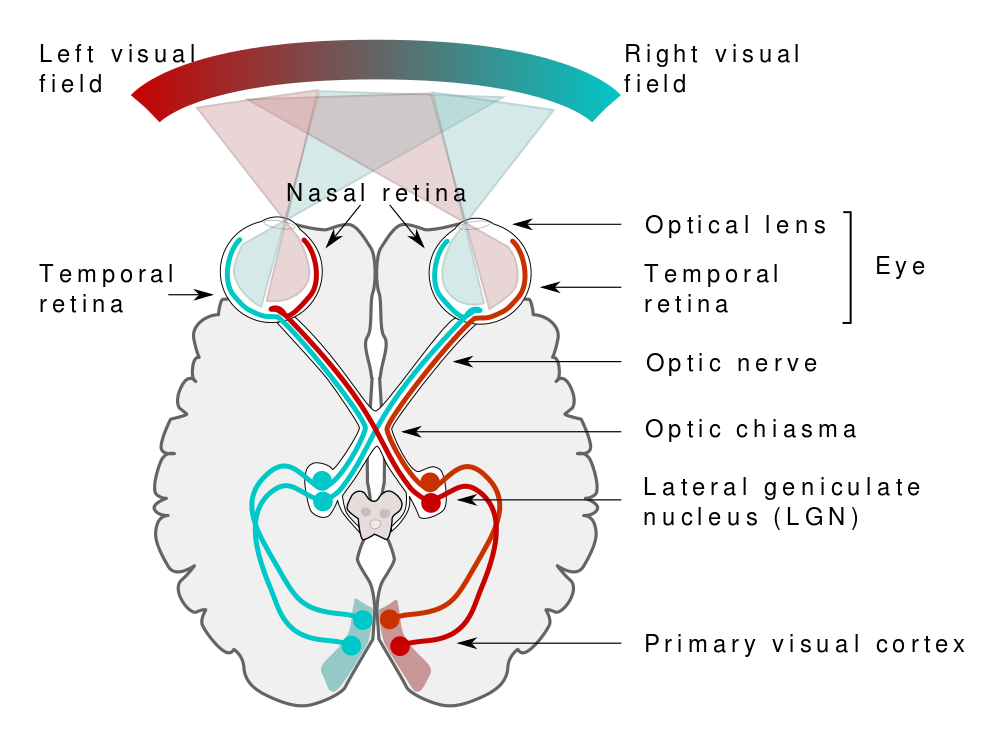
Visual Pathway. (Image by Miquel Perello Nieto (Own work) [CC BY-SA 4.0 , via Wikimedia Commons)
Blood Supply
Table: Blood Supply of the Visual Pathway
| Structure | Blood Supply |
|---|---|
| Optic nerve (retinal and orbital part) | Central retinal artery (branch of the ophthalmic artery) |
| Optic nerve (intracranial part) and optic chiasm | Ophthalmic artery, anterior cerebral and anterior communicating arteries |
| Optic tract | Posterior communicating artery and anterior choroidal artery |
| Lateral geniculate nucleus | Posterior cerebral artery and anterior choroidal artery |
| Optic radiations | Middle and posterior cerebral arteries, anterior choroidal artery |
| Visual cortex | Posterior cerebral artery (macular dually supplied by middle cerebral artery) |
Likely Causes of Disease or Injury
Damage to the optic nerve may be caused by:
- optic neuritis in multiple sclerosis or secondary to measles or mumps
- optic nerve compression secondary to orbital cellulitis, glaucoma or ocular tumours
- optic nerve toxicity secondary to ethambutol, methanol and ethylene glycol
- optic nerve damage secondary to orbital fracture or penetrating injury to the eye
- optic nerve ischaemia
Compression at the optic chiasm may occur due to:
- pituitary tumour
- craniopharyngioma
- meningioma
- optic glioma
- aneurysm of the internal carotid artery
Damage to the visual tract central to the optic chiasm may be caused by:
- cerebrovascular event
- brain abscess
- brain tumours
Common Clinical Effects
Table: Clinical Effects of Lesions of the Visual Pathway
| Site of Lesion | Visual Defect |
|---|---|
| Optic nerve | Ipsilateral visual loss |
| Optic chiasm | Bitemporal hemianopia |
| Optic tract | Contralateral homonymous hemianopia |
| Parietal upper optic radiation | Contralateral homonymous inferior quadrantanopia |
| Temporal lower optic radiation | Contralateral homonymous superior quadrantanopia |
| Occipital visual cortex | Contralateral homonymous hemianopia with macular sparing |
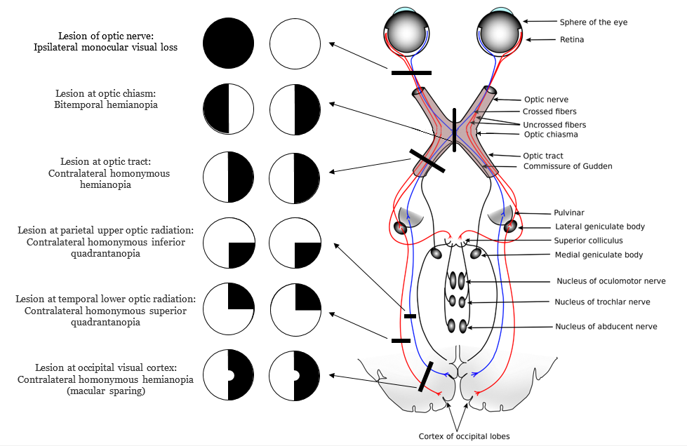
Clinical Effects of Lesions of the Visual Pathway. (Image modified by FRCEM Success. Original by KDS444 [Public domain], via Wikimedia Commons)
Report A Problem
Is there something wrong with this question? Let us know and we’ll fix it as soon as possible.
Loading Form...
- Biochemistry
- Blood Gases
- Haematology
| Biochemistry | Normal Value |
|---|---|
| Sodium | 135 – 145 mmol/l |
| Potassium | 3.0 – 4.5 mmol/l |
| Urea | 2.5 – 7.5 mmol/l |
| Glucose | 3.5 – 5.0 mmol/l |
| Creatinine | 35 – 135 μmol/l |
| Alanine Aminotransferase (ALT) | 5 – 35 U/l |
| Gamma-glutamyl Transferase (GGT) | < 65 U/l |
| Alkaline Phosphatase (ALP) | 30 – 135 U/l |
| Aspartate Aminotransferase (AST) | < 40 U/l |
| Total Protein | 60 – 80 g/l |
| Albumin | 35 – 50 g/l |
| Globulin | 2.4 – 3.5 g/dl |
| Amylase | < 70 U/l |
| Total Bilirubin | 3 – 17 μmol/l |
| Calcium | 2.1 – 2.5 mmol/l |
| Chloride | 95 – 105 mmol/l |
| Phosphate | 0.8 – 1.4 mmol/l |
| Haematology | Normal Value |
|---|---|
| Haemoglobin | 11.5 – 16.6 g/dl |
| White Blood Cells | 4.0 – 11.0 x 10⁹/l |
| Platelets | 150 – 450 x 109/l |
| MCV | 80 – 96 fl |
| MCHC | 32 – 36 g/dl |
| Neutrophils | 2.0 – 7.5 x 10⁹/l |
| Lymphocytes | 1.5 – 4.0 x 10⁹/l |
| Monocytes | 0.3 – 1.0 x 10⁹/l |
| Eosinophils | 0.1 – 0.5 x 10⁹/l |
| Basophils | < 0.2 x 10⁹/l |
| Reticulocytes | < 2% |
| Haematocrit | 0.35 – 0.49 |
| Red Cell Distribution Width | 11 – 15% |
| Blood Gases | Normal Value |
|---|---|
| pH | 7.35 – 7.45 |
| pO2 | 11 – 14 kPa |
| pCO2 | 4.5 – 6.0 kPa |
| Base Excess | -2 – +2 mmol/l |
| Bicarbonate | 24 – 30 mmol/l |
| Lactate | < 2 mmol/l |