
Anatomy
Head and Neck
The spinal cord is continuous superiorly with the:
Answer:
The spinal cord arises cranially as a continuation of the medulla oblongata at the foramen magnum.Spinal Cord
Anatomy / Central Nervous System
Last Updated: 27th June 2020
Extent
The spinal cord arises cranially as a continuation of the medulla oblongata at the foramen magnum. It then travels inferiorly within the vertebral canal, surrounded by the spinal meninges, before terminating as the conus medullaris. The filum terminale is an extension of pia mater that extends from the conus medullaris to the dural sac, and from the dural sac to the coccyx.
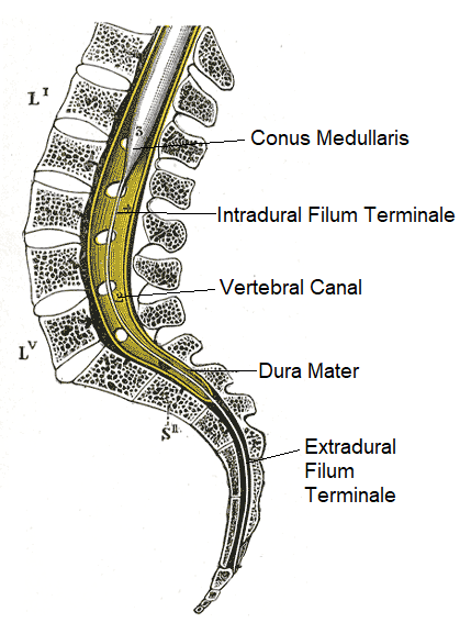
Extent of the Spinal Cord. (Image by Henry Vandyke Carter [Public domain], via Wikimedia Commons)
At birth, the conus medullaris lies at L3. By the age of 21, it sits at L1/L2, thus the spinal cord only occupies about 2/3s of the space available to it in the vertebral canal. The lower a nerve root, therefore, the more steeply it slopes down and the further it has to travel before gaining its intervertebral foramen. The lumbosacral nerve roots that arise from the end of the spinal cord and descend through the subarachnoid space to exit through their respective intervertebral or sacral foramina are bundled together forming the cauda equina.
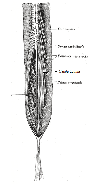
Cauda Equina. (Image modified by FRCEM Success. Original by Henry Vandyke Carter [Public domain], via Wikimedia Commons)
Enlargements
During the course of the spinal cord, there are two points of enlargement:
- The cervical enlargement is located in the region associated with the origins of spinal nerves C5 to T1 (at the C3 - T1 vertebral level).
- The lumbar enlargement is located in the region associated with the origins of spinal nerves L1 to S3 (at the T11 - L1 vertebral level).
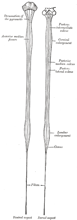
Enlargements of the Spinal Cord. (Image by Henry Vandyke Carter [Public domain], via Wikimedia Commons)
Internal anatomy
Internally the spinal cord has a small central canal (containing CSF) surrounded by grey and white matter. The central grey matter is rich in nerve cell bodies which are divided into three divisions (the anterior, posterior and lateral horns), which in cross-section form the characteristic H-shaped appearance in the central regions of the cord. The peripheral white matter surrounds the grey matter and is rich in myelinated nerve cell processes, which form large bundles or tracts that ascend and descend in the cord to other spinal cord levels or the brain. The white matter is divided bilaterally by sulci into three major divisions (the anterior, posterior and lateral columns).
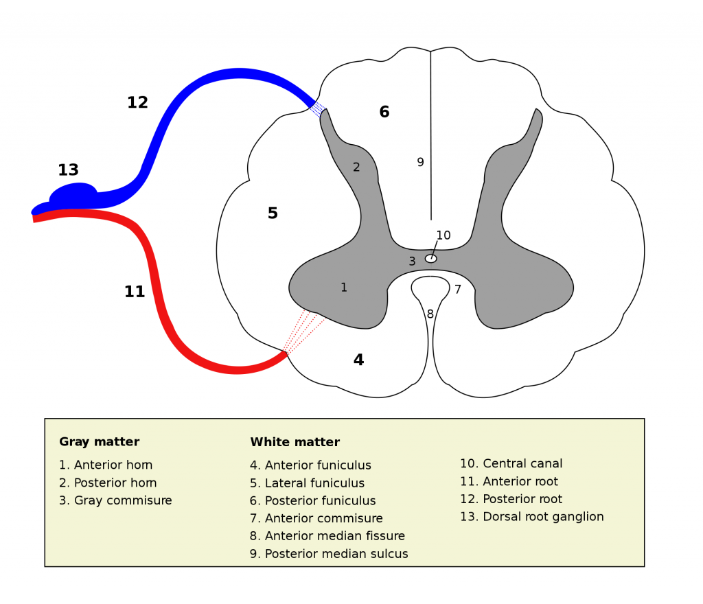
Internal Anatomy of the Spinal Cord. (Image by User:Polarlys (Own work) [GFDL , via Wikimedia Commons)
Blood Supply
The spinal cord is supplied primarily by branches of the vertebral artery:
- The single anterior spinal artery is formed from the left and right vertebral arteries and supplies the anterior two-thirds of the spinal cord, including the anterior and lateral horns and the anterior and lateral columns.
- The paired posterior spinal arteries arise from either the posterior inferior cerebellar artery or directly from the vertebral artery and supply the posterior third of the spinal cord including the posterior horns and columns.
Nerve Roots
The spinal nerves are formed by the union of posterior and anterior roots.
Spinal rootlets emerge from the spinal cord in the subarachnoid space and amalgamate shortly afterwards into roots. Anterior roots (containing motor nerve fibres) and posterior roots (containing the processes of sensory neurons) then emerge from their individual intervertebral foramina.
After invaginating the dura, the roots combine into mixed spinal nerves which divide into a posterior ramus (innervating intrinsic back muscles and an associated strip of skin on the back) and an anterior ramus (innervating the remaining muscles and skin of the trunk and limbs and the visceral organs).
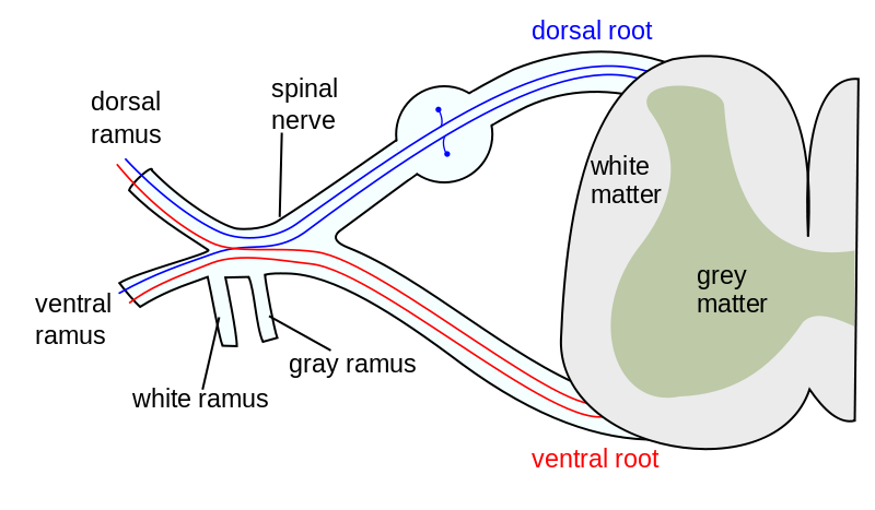
Spinal Nerve Roots. (Image by Mysid (original by Tristanb) [GFDL , via Wikimedia Commons)
Report A Problem
Is there something wrong with this question? Let us know and we’ll fix it as soon as possible.
Loading Form...
- Biochemistry
- Blood Gases
- Haematology
| Biochemistry | Normal Value |
|---|---|
| Sodium | 135 – 145 mmol/l |
| Potassium | 3.0 – 4.5 mmol/l |
| Urea | 2.5 – 7.5 mmol/l |
| Glucose | 3.5 – 5.0 mmol/l |
| Creatinine | 35 – 135 μmol/l |
| Alanine Aminotransferase (ALT) | 5 – 35 U/l |
| Gamma-glutamyl Transferase (GGT) | < 65 U/l |
| Alkaline Phosphatase (ALP) | 30 – 135 U/l |
| Aspartate Aminotransferase (AST) | < 40 U/l |
| Total Protein | 60 – 80 g/l |
| Albumin | 35 – 50 g/l |
| Globulin | 2.4 – 3.5 g/dl |
| Amylase | < 70 U/l |
| Total Bilirubin | 3 – 17 μmol/l |
| Calcium | 2.1 – 2.5 mmol/l |
| Chloride | 95 – 105 mmol/l |
| Phosphate | 0.8 – 1.4 mmol/l |
| Haematology | Normal Value |
|---|---|
| Haemoglobin | 11.5 – 16.6 g/dl |
| White Blood Cells | 4.0 – 11.0 x 10⁹/l |
| Platelets | 150 – 450 x 109/l |
| MCV | 80 – 96 fl |
| MCHC | 32 – 36 g/dl |
| Neutrophils | 2.0 – 7.5 x 10⁹/l |
| Lymphocytes | 1.5 – 4.0 x 10⁹/l |
| Monocytes | 0.3 – 1.0 x 10⁹/l |
| Eosinophils | 0.1 – 0.5 x 10⁹/l |
| Basophils | < 0.2 x 10⁹/l |
| Reticulocytes | < 2% |
| Haematocrit | 0.35 – 0.49 |
| Red Cell Distribution Width | 11 – 15% |
| Blood Gases | Normal Value |
|---|---|
| pH | 7.35 – 7.45 |
| pO2 | 11 – 14 kPa |
| pCO2 | 4.5 – 6.0 kPa |
| Base Excess | -2 – +2 mmol/l |
| Bicarbonate | 24 – 30 mmol/l |
| Lactate | < 2 mmol/l |

