Anatomy
Thorax
The oesophagus begins at which vertebral level:
Answer:
The oesophagus begins at the inferior border of the cricoid cartilage opposite vertebra C6, pierces the diaphragm at the esophageal hiatus at T10 and ends at the cardiac opening of the stomach opposite vertebra T11.Oesophagus (Thoracic)
Anatomy / Thorax / Oesophagus
Last Updated: 11th April 2019
The oesophagus is a muscular tube passing between the pharynx in the neck and the stomach in the abdomen.
Anatomical extent
The oesophagus begins at the inferior border of the cricoid cartilage opposite vertebra C6, pierces the diaphragm at the esophageal hiatus at T10 and ends at the cardiac opening of the stomach opposite vertebra T11.
Structure
The oesophagus consists of an internal circular and external longitudinal layer of muscle. The external longitudinal layer is comprised of different muscle types in each third of the oesophagus; superior third - voluntary striated muscle, middle third - voluntary striated and smooth muscle and inferior third - smooth muscle.
Relations
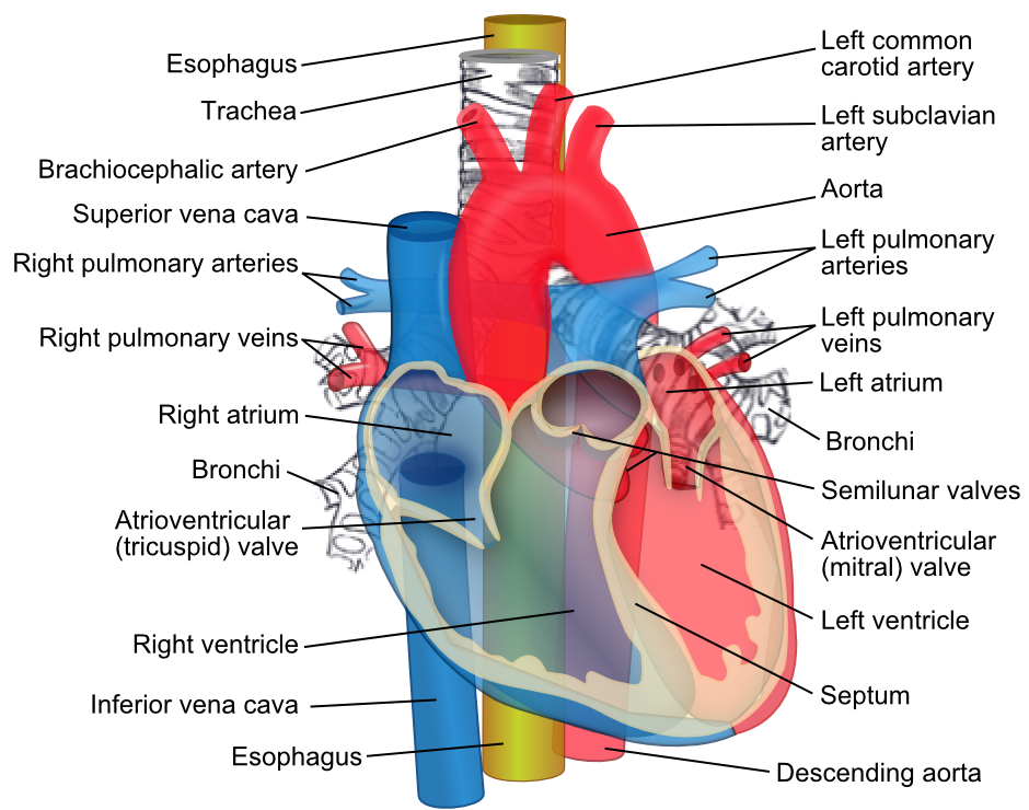
The oesophagus descends on the anterior aspect of the bodies of the vertebrae, immediately posterior to the trachea and the recurrent laryngeal nerves, generally in the midline position. The oesophagus is crossed anteriorly by the aortic arch. As it approaches the diaphragm, the oesophagus moves anteriorly and to the left, crossing from the right side of the thoracic aorta to eventually assume a position anterior to it, before passing through the diaphragm.
Posterior to the oesophagus, the thoracic duct is on the right side inferiorly but crosses to the left more superiorly (at T5). Other structures posteriorly include portions of the hemiazygos veins and the right posterior intercostal vessels.
Anterior to the oesophagus below the tracheal bifurcation are the right pulmonary artery and the left main bronchus. The oesophagus then passes immediately posterior to the left atrium, separated from it only by pericardium. Inferior to the left atrium, the oesophagus is related to the diaphragm.

Relations of the Oesophagus. (Image by ZooFari [CC BY-SA 3.0 , via Wikimedia Commons)
Points of constriction
As the oesophagus is a flexible muscular tube, it is compressed by surrounding structures at four locations:
- 1) the junction of the oesophagus with the pharynx in the neck (upper oesophageal sphincter formed by the cricopharyngeus muscle)
- 2) in the superior mediastinum where the oesophagus is crossed by the arch of the aorta
- 3) in the posterior mediastinum where the oesophagus is compressed by the left main bronchus
- 4) in the posterior mediastinum at the oesophageal hiatus in the diaphragm.
Clinically these compressions are important when passing instruments or assessing swallowed objects.
Innervation
Oesophageal innervation is complex, oesophageal branches arise from the vagus nerves and sympathetic trunks. The visceral afferents that pass through the sympathetic trunks and the splanchnic nerves are the primary participants in detection of oesophageal pain and transmission of this information to various levels of the central nervous system.
Report A Problem
Is there something wrong with this question? Let us know and we’ll fix it as soon as possible.
Loading Form...
- Biochemistry
- Blood Gases
- Haematology
| Biochemistry | Normal Value |
|---|---|
| Sodium | 135 – 145 mmol/l |
| Potassium | 3.0 – 4.5 mmol/l |
| Urea | 2.5 – 7.5 mmol/l |
| Glucose | 3.5 – 5.0 mmol/l |
| Creatinine | 35 – 135 μmol/l |
| Alanine Aminotransferase (ALT) | 5 – 35 U/l |
| Gamma-glutamyl Transferase (GGT) | < 65 U/l |
| Alkaline Phosphatase (ALP) | 30 – 135 U/l |
| Aspartate Aminotransferase (AST) | < 40 U/l |
| Total Protein | 60 – 80 g/l |
| Albumin | 35 – 50 g/l |
| Globulin | 2.4 – 3.5 g/dl |
| Amylase | < 70 U/l |
| Total Bilirubin | 3 – 17 μmol/l |
| Calcium | 2.1 – 2.5 mmol/l |
| Chloride | 95 – 105 mmol/l |
| Phosphate | 0.8 – 1.4 mmol/l |
| Haematology | Normal Value |
|---|---|
| Haemoglobin | 11.5 – 16.6 g/dl |
| White Blood Cells | 4.0 – 11.0 x 10⁹/l |
| Platelets | 150 – 450 x 109/l |
| MCV | 80 – 96 fl |
| MCHC | 32 – 36 g/dl |
| Neutrophils | 2.0 – 7.5 x 10⁹/l |
| Lymphocytes | 1.5 – 4.0 x 10⁹/l |
| Monocytes | 0.3 – 1.0 x 10⁹/l |
| Eosinophils | 0.1 – 0.5 x 10⁹/l |
| Basophils | < 0.2 x 10⁹/l |
| Reticulocytes | < 2% |
| Haematocrit | 0.35 – 0.49 |
| Red Cell Distribution Width | 11 – 15% |
| Blood Gases | Normal Value |
|---|---|
| pH | 7.35 – 7.45 |
| pO2 | 11 – 14 kPa |
| pCO2 | 4.5 – 6.0 kPa |
| Base Excess | -2 – +2 mmol/l |
| Bicarbonate | 24 – 30 mmol/l |
| Lactate | < 2 mmol/l |

