Pathology
Haematology
Which of the following laboratory findings is NOT typical of von Willebrand disease (VWD):
Answer:
Laboratory findings typically show (although this varies depending on VWD type):- Abnormal PFA-100 test
- Low factor VIII levels (if low a factor VIII/VWF binding assay is performed)
- Prolonged APTT (or normal)
- Normal PT
- Low VWF levels
- Defective platelet aggregation
- Normal platelet count
Inherited Coagulation Disorders
Pathology / Haematology / Coagulation Disorders
Last Updated: 24th April 2019
Hereditary deficiencies of each of the coagulation factors have been described. Haemophilia A (factor VIII) deficiency, haemophilia B (Christmas disease, factor IX deficiency) and von Willebrand disease (VWD) are the most frequent; the others are rarer.
Haemophilia A
Haemophilia A is the most common of the hereditary clotting factor deficiencies. The inheritance is sex-linked but up to one-third of patients have no family history and these cases result from recent mutation. The defect is an absence or low level of plasma factor VIII.
Inheritance:
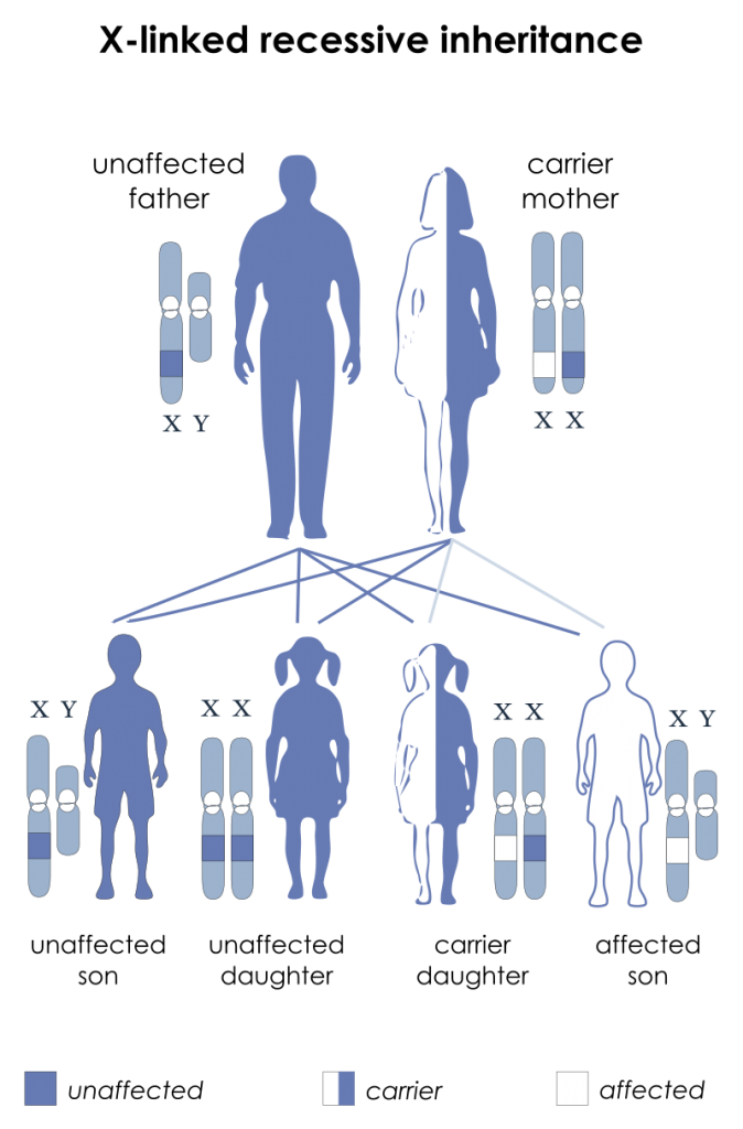
The inheritance is X-linked recessive; therefore (assuming the other parent is a non-carrier):
- Sons of a female carrier have a 50% chance of developing the disease
- Daughters of a female carrier have a 50% chance of being a carrier
- All sons of a male haemophiliac will be unaffected
- All daughters of a male haemophiliac will be a carrier

X-linked Recessive Inheritance Pattern. (Image by National Institutes of Health derivative work: Drsrisenthil [Public domain])
Infants may develop profuse post-circumcision haemorrhage or joint and soft tissue bleeds and excessive bruising when they start to become active. Recurrent painful haemarthroses and muscle haematomas dominate the clinical course of severely affected patients and if inadequately treated, lead to progressive joint deformity and disability. Local pressure can cause entrapment neuropathy or ischaemic necrosis. Prolonged bleeding occurs after dental extractions or post-trauma. Spontaneous haematuria and gastrointestinal haemorrhage may occur. The clinical severity of the disease correlates inversely with the factor VIII level. Operative and post-traumatic haemorrhage are life-threatening both in severely and mildly affected patients. Haemophilic pseudotumours are large encapsulated haematomas with progressive cystic swelling from repeated haemorrhage. Due to blood product transfusions, many patients were infected with HIV and hepatitis C during the 1980s.
Laboratory Findings:
- Prolonged APTT
- Normal PT
- Low Factor VIII
Haemophilia B (Christmas Disease)
Haemophilia B is due to a factor IX deficiency. The inheritance and clinical features are identical to that of haemophilia A. The two disorders can only be distinguished by specific coagulation factor assays. The incidence is one-fifth of that of haemophilia A. Laboratory findings demonstrate prolonged APTT, normal PT and low factor IX.
Von Willebrand Disease
Von Willebrand disease (VWD) results from either a reduced level or abnormal functioning of von Willebrand factor (VWF) resulting from a wide variety of genetic mutations.
VWF is produced both in endothelial cells and megakaryocytes. VWF has two roles: it promotes platelet adhesion to subendothelium and to each other at high shear rates and it is the carrier molecule for factor VIII, protecting it from premature destruction. The latter property explains the reduced factor VIII levels found in VWD.
VWD is the most common inherited bleeding disorder. Usually the inheritance is autosomal dominant, and the severity of the bleeding is highly variable. Women are more badly affected than men at a given VWF level. Typically there is mucous membrane bleeding (e.g. epistaxis, menorrhagia), excessive blood loss from superficial cuts and abrasions, and operative and post-traumatic haemorrhage. Haemarthroses and muscle haematomas are rare.
Laboratory findings typically show (although again, this varies depending on VWD type):
- Abnormal PFA-100 test
- Low factor VIII levels (if low a factor VIII/VWF binding assay is performed)
- Prolonged APTT (or normal)
- Normal PT
- Low VWF levels
- Defective platelet aggregation
- Normal platelet count
Report A Problem
Is there something wrong with this question? Let us know and we’ll fix it as soon as possible.
Loading Form...
- Biochemistry
- Blood Gases
- Haematology
| Biochemistry | Normal Value |
|---|---|
| Sodium | 135 – 145 mmol/l |
| Potassium | 3.0 – 4.5 mmol/l |
| Urea | 2.5 – 7.5 mmol/l |
| Glucose | 3.5 – 5.0 mmol/l |
| Creatinine | 35 – 135 μmol/l |
| Alanine Aminotransferase (ALT) | 5 – 35 U/l |
| Gamma-glutamyl Transferase (GGT) | < 65 U/l |
| Alkaline Phosphatase (ALP) | 30 – 135 U/l |
| Aspartate Aminotransferase (AST) | < 40 U/l |
| Total Protein | 60 – 80 g/l |
| Albumin | 35 – 50 g/l |
| Globulin | 2.4 – 3.5 g/dl |
| Amylase | < 70 U/l |
| Total Bilirubin | 3 – 17 μmol/l |
| Calcium | 2.1 – 2.5 mmol/l |
| Chloride | 95 – 105 mmol/l |
| Phosphate | 0.8 – 1.4 mmol/l |
| Haematology | Normal Value |
|---|---|
| Haemoglobin | 11.5 – 16.6 g/dl |
| White Blood Cells | 4.0 – 11.0 x 10⁹/l |
| Platelets | 150 – 450 x 109/l |
| MCV | 80 – 96 fl |
| MCHC | 32 – 36 g/dl |
| Neutrophils | 2.0 – 7.5 x 10⁹/l |
| Lymphocytes | 1.5 – 4.0 x 10⁹/l |
| Monocytes | 0.3 – 1.0 x 10⁹/l |
| Eosinophils | 0.1 – 0.5 x 10⁹/l |
| Basophils | < 0.2 x 10⁹/l |
| Reticulocytes | < 2% |
| Haematocrit | 0.35 – 0.49 |
| Red Cell Distribution Width | 11 – 15% |
| Blood Gases | Normal Value |
|---|---|
| pH | 7.35 – 7.45 |
| pO2 | 11 – 14 kPa |
| pCO2 | 4.5 – 6.0 kPa |
| Base Excess | -2 – +2 mmol/l |
| Bicarbonate | 24 – 30 mmol/l |
| Lactate | < 2 mmol/l |

