Anatomy
Abdomen
The liver is divided into the left and right lobes by which of the following structures:
Answer:
The liver is divided into the left and right lobe by the falciform ligament.Liver
Anatomy / Abdomen / Liver and Biliary Tract
Last Updated: 22nd November 2020
The liver is the largest visceral organ in the body and is primarily located in the right hypochondrium and epigastric region, extending into the left hypochondrium.
Surface Markings
Most of the liver is under the right dome of the diaphragm and deep to the lower thoracic wall. The inferior margin of the liver can be palpated descending below the right costal margin on deep inspiration.
The inferior margin is indicated by a line that joins points at the right tenth costal cartilage in the midaxillary line, the tip of the right ninth costal cartilage, the transpyloric plane in the midline, the tip of the left eighth costal cartilage and the left fifth rib in the midclavicular line.
The superior margin is indicated by a line that joins a point on the left fifth rib at the midclavicular line to a corresponding point on the right side. The superior margin is concave in its central part and crosses behind the xiphisternal joint.

Surface Markings of the Liver. (Image by Henry Vandyke Carter [Public domain], via Wikimedia Commons)
Anatomical Surfaces
The liver has a diaphragmatic surface in the anterior, superior, and posterior directions and a visceral surface in the inferior direction.
The diaphragmatic surface of the liver is related anteriorly to the anterior abdominal wall and rib cage and superiorly to the diaphragm.
The visceral surface of the liver is related to the oesophagus, right anterior part of the stomach, superior part of the duodenum, lesser omentum, gallbladder, right colic flexure, right transverse colon, right kidney and right adrenal gland.
![By Henry Vandyke Carter [Public domain], via Wikimedia Commons](https://mrcemsuccess.com/wp-content/uploads/2017/02/Liver2.png)
Diaphragmatic Surface of the Liver. (Image by Henry Vandyke Carter [Public domain], via Wikimedia Commons)

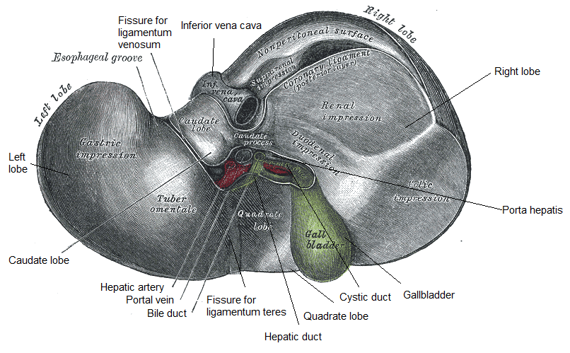
Inferior Surface of the Liver. (Image modified by FRCEM Success. Original by Henry Vandyke Carter [Public domain], via Wikimedia Commons)
Lobes
There are four anatomical lobes to the liver.
- The liver is divided into the left and right lobe by falciform ligament.
- The caudate lobe sits between the fissure for the ligamentum venosum and the inferior vena cava.
- The quadrate lobe is located between the gallbladder and the fissure for the ligamentum teres.
The porta hepatis is the central intraperitoneal fissure of the liver that separates the caudate and the quadrate lobes. The porta hepatis serves as the point of entry into the liver for the hepatic arteries and portal vein, and the exit point for the hepatic ducts.
Functional Anatomy
Microscopically, hepatocytes are arranged into lobules which are the functional units of the liver. Each lobule is hexagonal-shaped, and is drained by a venule in its centre, called a central vein (which drains to the hepatic vein).
At the periphery of the hexagon are three structures collect0vely known as the portal triad, comprising:
- a portal arteriole (a branch of the hepatic artery),
- a portal venule (a branch of the hepatic portal vein),
- a bile duct.
The portal triad also contains lymphatic vessels and vagal parasympathetic nerve fibres.
The liver sinusoids serve as a location for mixing of the oxygen-rich blood from the hepatic artery and the nutrient-rich blood from the portal vein.

Functional Anatomy of the Liver (Image by OpenStax College [CC BY 3.0 , via Wikimedia Commons)
Blood supply
The liver has a unique dual blood supply. The arterial supply to the liver is from the left and right hepatic arteries derived from the hepatic artery proper, a branch of the common hepatic artery from the coeliac trunk.
The hepatic portal vein (formed from the union of the superior mesenteric and splenic vein posterior to the neck of the pancreas at the level of vertebra L2) supplies the liver with deoxygenated blood carrying nutrients absorbed from the small intestine.
Venous drainage of the liver is achieved through three hepatic veins, which return the venous blood to the inferior vena cava just inferior to the diaphragm.
Lymphatic drainage
The lymphatic vessels of the liver drain into hepatic lymph nodes which empty in the coeliac lymph nodes.
Report A Problem
Is there something wrong with this question? Let us know and we’ll fix it as soon as possible.
Loading Form...
- Biochemistry
- Blood Gases
- Haematology
| Biochemistry | Normal Value |
|---|---|
| Sodium | 135 – 145 mmol/l |
| Potassium | 3.0 – 4.5 mmol/l |
| Urea | 2.5 – 7.5 mmol/l |
| Glucose | 3.5 – 5.0 mmol/l |
| Creatinine | 35 – 135 μmol/l |
| Alanine Aminotransferase (ALT) | 5 – 35 U/l |
| Gamma-glutamyl Transferase (GGT) | < 65 U/l |
| Alkaline Phosphatase (ALP) | 30 – 135 U/l |
| Aspartate Aminotransferase (AST) | < 40 U/l |
| Total Protein | 60 – 80 g/l |
| Albumin | 35 – 50 g/l |
| Globulin | 2.4 – 3.5 g/dl |
| Amylase | < 70 U/l |
| Total Bilirubin | 3 – 17 μmol/l |
| Calcium | 2.1 – 2.5 mmol/l |
| Chloride | 95 – 105 mmol/l |
| Phosphate | 0.8 – 1.4 mmol/l |
| Haematology | Normal Value |
|---|---|
| Haemoglobin | 11.5 – 16.6 g/dl |
| White Blood Cells | 4.0 – 11.0 x 10⁹/l |
| Platelets | 150 – 450 x 109/l |
| MCV | 80 – 96 fl |
| MCHC | 32 – 36 g/dl |
| Neutrophils | 2.0 – 7.5 x 10⁹/l |
| Lymphocytes | 1.5 – 4.0 x 10⁹/l |
| Monocytes | 0.3 – 1.0 x 10⁹/l |
| Eosinophils | 0.1 – 0.5 x 10⁹/l |
| Basophils | < 0.2 x 10⁹/l |
| Reticulocytes | < 2% |
| Haematocrit | 0.35 – 0.49 |
| Red Cell Distribution Width | 11 – 15% |
| Blood Gases | Normal Value |
|---|---|
| pH | 7.35 – 7.45 |
| pO2 | 11 – 14 kPa |
| pCO2 | 4.5 – 6.0 kPa |
| Base Excess | -2 – +2 mmol/l |
| Bicarbonate | 24 – 30 mmol/l |
| Lactate | < 2 mmol/l |

