

Physiology
Cardiovascular
In the ventricular ejection phase of the cardiac cycle, all of the following statements are true EXCEPT for:
Answer:
When the ventricular pressure exceeds that in the pulmonary artery and the aorta, the semilunar valves open and blood is ejected, initially rapidly (rapid ejection phase) and then more slowly (reduced ejection phase). The AV valves which closed at the beginning of isovolumetric contraction, remain closed in the ventricular ejection phase. Atrial pressure initially decreases as the atrial base is pulled downward during ejection, expanding the atrial chamber (the x descent of the JVP waveform). During the second half of ejection, the ventricles stop actively contracting, the ventricular pressure starts to decrease and the muscle starts to repolarise; this causes the T wave on the ECG, which marks the end of both ventricular contraction and rapid ventricular ejection.Pressures, Volumes and Key Events in Cardiac Cycle
Physiology / Cardiovascular / Cardiac Cycle
Last Updated: 31st January 2022
The cardiac cycle describes the events that occur during one beat of the heart.
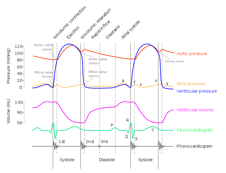
Cardiac Cycle. (Image modified by FRCEM Success. Original by DanielChangMD revised original work of DestinyQx; Redrawn as SVG by xavax [CC BY-SA 2.5 (https://creativecommons.org/licenses/by-sa/2.5)], via Wikimedia Commons)
Diastole: Atrial Systole (AV valves open, Semilunar valves closed)
Atrial depolarisation causes the P wave on the ECG and initiates atrial contraction (atrial repolarisation is too diffuse to be seen on the ECG).
As the atria contract, the atrial pressure increases which forces more blood flow across the open AV valves, leading to rapid flow of blood into the ventricles. There are no valves between the veins and atria and atrial systole causes a small pressure rise in the great veins (the a wave on the JVP waveform).
At rest, atrial contraction only contributes the last 15 - 20% of the final ventricular volume, as most of the ventricular filling has occurred passively in diastole due to venous pressure. The proportion of atrial contribution increases with heart rate as diastole shortens and there is less time for passive ventricular filling.
The end-diastolic volume (EDV) is usually about 120 - 140 mL, and the end-diastolic pressure is less than 10 mmHg (and higher in the left ventricle than the right due to the thicker and therefore stiffer left ventricle).
In ventricular hypertrophy, filling of the 'stiff' ventricle by atrial systole causes a fourth heart sound, which is not audible in normal adults.
Systole: Isovolumetric Contraction (All valves closed)
Ventricular depolarisation causes the QRS complex on the ECG, and triggers excitation-contraction coupling and myocyte contraction.
The ventricular pressure rises sharply during contraction and the AV valves close as soon as this is greater than the atrial pressure (causing the first heart sound). Because the mitral valve closes before the tricuspid valve, the first heart sound may be split.
For a short period, as the forces are developing, both the AV and the semilunar valves are closed as the ventricular pressure is still less than that in the pulmonary artery and aorta, and no ejection occurs. This is isovolumetric contraction.
The increasing pressure makes the AV valves bulge into the atria, causing a small atrial pressure wave (the c wave of the JVP waveform).
Systole: Ventricular Ejection (Semilunar valves open, AV valves closed)
When the ventricular pressure exceeds that in the pulmonary artery and the aorta, the semilunar valves open and blood is ejected, initially rapidly (rapid ejection phase) and then more slowly (reduced ejection phase).
Atrial pressure initially decreases as the atrial base is pulled downward during ejection, expanding the atrial chamber (the x descent of the JVP waveform). Atrial filling begins in the rapid ejection phase and continues during the reduced ejection phase and atrial pressure begins to rise (the v wave of the JVP waveform).
During the second half of ejection, the ventricles stop actively contracting, the ventricular pressure starts to decrease and the muscle starts to repolarise; this causes the T wave on the ECG, which marks the end of both ventricular contraction and rapid ventricular ejection.
The ventricular pressure during the reduced ejection phase begins to decrease. Aortic pressure also decreases because of the runoff of blood from large arteries into smaller arteries. The ventricular pressure falls slightly below that in the aorta, but initially blood continues to flow out of the ventricle because of momentum; eventually the ventricular pressure falls sufficiently and the semilunar valves close.
The amount of blood ejected is the stroke volume (SV), and is usually about 70 mL (therefore about 50 mL is left; this is the end-systolic volume). The proportion of EDV that is ejected (i.e. the SV/EDV) is the ejection fraction and this is normally about 0.6.
Diastole: Isovolumetric Relaxation (All valves closed)
Closure of the semilunar valves causes a small increase in aortic pressure (the dicrotic notch on the arterial waveform), and the second heart sound. Inspiration delays closure of the pulmonary valve and thus causes splitting of the second heart sound.
Immediately after the closure of the semilunar valves, the ventricles rapidly relax and ventricular pressure decreases rapidly but the AV valves remain closed as initially the ventricular pressure is still greater than atrial pressure. This is isovolumetric relaxation.
Atrial pressure continues to rise because of venous return, with the v wave of the JVP waveform peaking during this phase. As the ventricles continue to relax, the ventricular pressure falls below that of the atrial pressure and the AV valves open.
Diastole: Ventricular Filling (AV valves open, Semilunar valves closed)
When the AV valves open, the atrial pressure falls (the y descent of the JVP waveform) and the ventricles refill, initially rapidly (the rapid filling phase) and then more slowly as the ventricles expand, become less compliant, and ventricular pressures rise (the reduced filling phase). Rapid flow of blood from the atria into the ventricles causes the third heart sound, which is normal in children but, in adults, is associated with disease such as ventricular dilation.
Diastole is usually twice the length of systole at rest, but decreases with increased heart rate. During systole, contraction of the ventricles compresses the coronary arteries and suppresses blood flow. This is particularly evident in the left ventricle, where during systole the ventricular pressure is the same as or greater than that in the arteries and as a result more than 85% of left ventricular perfusion occurs during diastole. This becomes a problem if the heart rate is increased as the diastolic interval is shorter and can result in ischaemia.
Valve Opening/Closure during Cardiac Cycle
| Cardiac Cycle Phase | Atrioventricular Valves | Semilunar Valves |
|---|---|---|
| Atrial systole | Open (atrial pressure > ventricular pressure) | Closed (arterial pressure > ventricular pressure) |
| Isovolumetric contraction | Closed (ventricular pressure > atrial pressure) | Closed (arterial pressure > ventricular pressure) |
| Ventricular ejection | Closed (ventricular pressure > atrial pressure) | Open (ventricular pressure > arterial pressure) |
| Isovolumetric relaxation | Closed (ventricular pressure > atrial pressure) | Closed (arterial pressure > ventricular pressure) |
| Ventricular filling | Open (atrial pressure > ventricular pressure) | Closed (arterial pressure > ventricular pressure) |
JVP Waveform during Cardiac Cycle
| JVP waveform | Phase of Cardiac Cycle | Physiology |
|---|---|---|
| a wave | Atrial systole (end diastole) | Occurs due to right atrial contraction |
| c wave | Isovolumetric contraction (early systole) | Occurs due to the bulging of the tricuspid valve into the right atrium during right isovolumetric ventricular contraction |
| x descent | Rapid ventricular ejection (mid systole) | Occurs due to a combination of right atrial relaxation, the downward displacement of the tricuspid valve during right ventricular contraction, and the ejection of blood from both the ventricles |
| v wave | Ventricular ejection and isovolumetric relaxation (late systole) | Occurs due to right atrial filling from venous return |
| y descent | Ventricular filling (early diastole) | Occurs due to opening of the tricuspid valve and the subsequent rapid inflow of blood from the right atrium to the right ventricle |
Heart Sounds during Cardiac Cycle
| Heart Sound | Phase of Cardiac Cycle | Mechanical Event |
|---|---|---|
| First heart sound | Start of systole | Caused by closure of the atrioventricular (mitral and tricuspid) valves |
| Second heart sound | End of systole | Caused by closure of the semilunar (aortic and pulmonary) valves |
| Third heart sound | Early diastole | Caused by rapid flow of blood from the atria into the ventricles during the ventricular filling phase |
| Fourth heart sound | Late diastole | Caused by filling of an abnormally stiff ventricle in atrial systole |
ECG Deflections during Cardiac Cycle
| ECG | Event |
|---|---|
| P wave | Atrial depolarisation |
| QRS complex | Ventricular depolarisation |
| T wave | Ventricular repolarisation |
Report A Problem
Is there something wrong with this question? Let us know and we’ll fix it as soon as possible.
Loading Form...
- Biochemistry
- Blood Gases
- Haematology
| Biochemistry | Normal Value |
|---|---|
| Sodium | 135 – 145 mmol/l |
| Potassium | 3.0 – 4.5 mmol/l |
| Urea | 2.5 – 7.5 mmol/l |
| Glucose | 3.5 – 5.0 mmol/l |
| Creatinine | 35 – 135 μmol/l |
| Alanine Aminotransferase (ALT) | 5 – 35 U/l |
| Gamma-glutamyl Transferase (GGT) | < 65 U/l |
| Alkaline Phosphatase (ALP) | 30 – 135 U/l |
| Aspartate Aminotransferase (AST) | < 40 U/l |
| Total Protein | 60 – 80 g/l |
| Albumin | 35 – 50 g/l |
| Globulin | 2.4 – 3.5 g/dl |
| Amylase | < 70 U/l |
| Total Bilirubin | 3 – 17 μmol/l |
| Calcium | 2.1 – 2.5 mmol/l |
| Chloride | 95 – 105 mmol/l |
| Phosphate | 0.8 – 1.4 mmol/l |
| Haematology | Normal Value |
|---|---|
| Haemoglobin | 11.5 – 16.6 g/dl |
| White Blood Cells | 4.0 – 11.0 x 10⁹/l |
| Platelets | 150 – 450 x 109/l |
| MCV | 80 – 96 fl |
| MCHC | 32 – 36 g/dl |
| Neutrophils | 2.0 – 7.5 x 10⁹/l |
| Lymphocytes | 1.5 – 4.0 x 10⁹/l |
| Monocytes | 0.3 – 1.0 x 10⁹/l |
| Eosinophils | 0.1 – 0.5 x 10⁹/l |
| Basophils | < 0.2 x 10⁹/l |
| Reticulocytes | < 2% |
| Haematocrit | 0.35 – 0.49 |
| Red Cell Distribution Width | 11 – 15% |
| Blood Gases | Normal Value |
|---|---|
| pH | 7.35 – 7.45 |
| pO2 | 11 – 14 kPa |
| pCO2 | 4.5 – 6.0 kPa |
| Base Excess | -2 – +2 mmol/l |
| Bicarbonate | 24 – 30 mmol/l |
| Lactate | < 2 mmol/l |