Revision Resources
Recent Posts View All
December FOAMed
Lemierre’s Syndrome

A 23-year-old male presents for severe throat pain and cough. He states that his neck hurts, noticing that the left side of his neck is red and painful. Review of systems is remarkable for a recent bout of pharyngitis that initially improved.
Vital signs include BP 91/49, HR 130, T 102.2 temporal, RR 25, SpO2 91% on room air. He appears toxic. The ENT exam reveals a midline uvula; soft mouth floor; prominent generalized cervical, submandibular, and submental swelling with corresponding lymphadenopathy; but no voice changes or difficulty tolerating secretions. His neck is red and tender, with mild swelling overlying the left side of neck and a painful tracheal rock. He is tachycardic and tachypneic, with multiple areas of wheezes and rhonchi.
What is the most likely diagnosis?
Congenital Cardiac Lesions

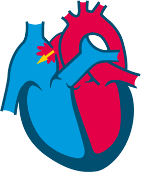
There are generally 3 main congenital heart diseases presentations that young children between ages of 0 days to about 3 months will present with. Often times you will see experts split these congenital heart lesions into 2 categories: Cyanotic vs Acyanotic lesions. Either is an acceptable approach as long as you split the acyanotic lesions into 2 categories as their presentations vary widely as I will describe.
Simple Febrile Seizures

An 18-month-old male presents to the ED with his parents for evaluation of seizure. His mother is at bedside providing the history. She states the patient had a runny nose beginning yesterday. This morning, he woke up and “felt warm” when he woke up but she did not take his temperature. About 20 minutes later, she noticed he was “out of it” and was “shaking all over.” She panicked and called 911 immediately. She thinks the shaking lasted about 2-4 minutes. By the time paramedics arrived, he had stopped shaking but was very sleepy. The patient’s blood glucose was 95. His temperature was 102 F (38.9 C) and they transported on pulse oximetry, which was normal en route. The patient is back to his baseline now and playing.
Testicular Torsion

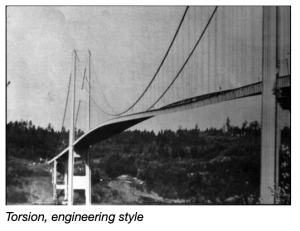
A 27-year-old male presents to the ED with left groin pain for the past 2 days. He had a similar episode lasting 30 minutes 3 days previously. Exam shows marked tenderness in the left inguinal region. Testicle is documented as normal size and non-tender. A radiologist is consulted and suggests a CT scan of the abdomen to rule out hernia, which shows a thickened left spermatic cord. A diagnosis of epidydimitis is made, and he is discharged. He returns 26 hours later with a swollen testicle and is diagnosed with torsion. The testicle cannot be salvaged. The patient consults an attorney who has an expert review the record.
Penetrating Wounds to the Groin

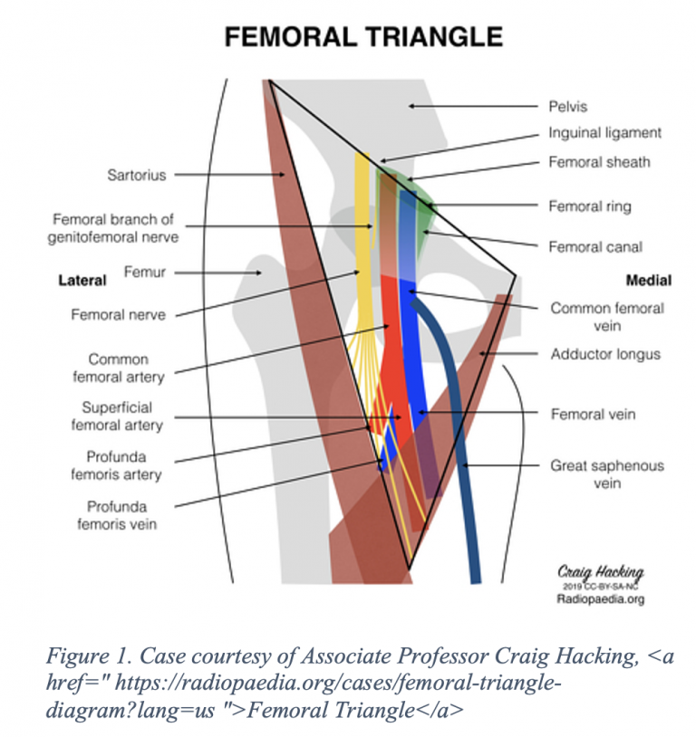
A 23-year-old male presents with a single stab wound to the left groin. The blade was estimated to be 3-4 inches long. The wound is 2 cm wide with dark blood slowly oozing from the site. Vital signs as follows: BP 153/75, HR 92, RR 12, SpO2 98%. What is the next step in management? How would you evaluate for this patient’s injuries?
Are you sure you wish to end this session?

